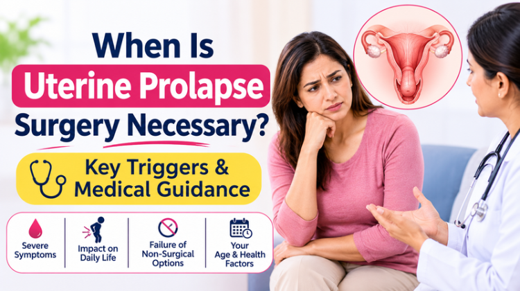
When Is Uterine Prolapse Surgery Necessary? Key Triggers & Medical Guidance
Pelvic floor disorders impact millions of women worldwide, yet open discussions regarding pelvic organ displacement remain surprisingly limited. Among these structural conditions, uterine descent represents a significant medical challenge that alters physical comfort, urinary function, and daily quality of life. Understanding when conservative management ceases to be effective and when uterine prolapse surgery becomes essential empowers patients to make informed decisions about their pelvic health.
The pelvic floor functions as a muscular hammock that supports the pelvic organs—including the uterus, bladder, and rectum. When childbirth, hormonal shifts during menopause, chronic intra-abdominal pressure, or aging weaken these supportive ligaments and fascia, the uterus can descend into or beyond the vaginal canal. While early-stage prolapse often responds well to conservative therapies, advanced organ displacement typically requires structural surgical repair to restore pelvic anatomy and functional well-being.
Understanding Uterine Prolapse and Structural Staging
To evaluate whether surgical intervention is required, clinical specialists categorize uterine descent into four standardized anatomical stages using systems such as the Pelvic Organ Prolapse Quantification (POP-Q) scale. Staging determines how far the organ has moved from its natural position:
-
Stage 1 (Mild Prolapse): The cervix descends into the upper half of the vaginal canal but remains well above the vaginal opening.
-
Stage 2 (Moderate Prolapse): The cervix or lower uterine segment extends close to the hymenal ring or vaginal opening.
-
Stage 3 (Severe Prolapse): The cervix and body of the uterus protrude significantly beyond the vaginal introitus.
-
Stage 4 (Complete Procidentia): The entire uterus lies outside the body, causing total eversion of the vaginal walls.
While Stage 1 and Stage 2 prolapse can often be managed through non-surgical protocols, Stage 3 and Stage 4 descent almost universally necessitate uterine prolapse surgery to prevent progressive tissue breakdown and severe visceral dysfunction.
Key Triggers: When Is Uterine Prolapse Surgery Necessary?
Determining the exact point at which conservative measures are no longer sufficient involves evaluating physical symptoms, functional impairments, and anatomical progression. Healthcare providers look for specific clinical triggers before recommending surgical reconstruction.
1. Failure of Non-Surgical and Conservative Treatments
The primary line of defense for mild to moderate pelvic floor weakness includes targeted pelvic floor physical therapy (Kegel exercises), weight management, and the fitting of a vaginal pessary (a removable silicone support device). However, surgery is strongly indicated when:
-
A pessary fails to remain in position due to severe floor relaxation.
-
The patient develops persistent vaginal mucosal erosions, ulcerations, or allergic reactions to pessary materials.
-
Pelvic floor exercises show no clinical halt in the physical descent of the organs.
2. Severe Bulging and Visible Tissue Protrusion
When pelvic organs drop past the vaginal opening, constant friction against undergarments causes raw, painful mucosal changes. Tissue protrusion leads to:
-
Recurrent vaginal bleeding or discharge secondary to mechanical ulceration.
-
Inability to sit, walk, or perform daily physical tasks comfortably.
-
Physical obstruction that interferes with basic mobility and bodily functions.
In such cases, uterine prolapse surgery serves as the definitive solution to elevate and secure the displaced anatomy.
3. Progressive Bladder and Urinary Dysfunction
Because the bladder is anatomically tethered to the front wall of the uterus and vagina, severe uterine descent pulls the bladder out of alignment (cystocele). Surgical intervention becomes medically necessary when urinary complications arise, such as:
-
Urinary Retention: The displaced uterus kinks the urethra, preventing full bladder drainage.
-
Recurrent Urinary Tract Infections (UTIs): Stagnant urine remaining in the bladder acts as a reservoir for bacterial growth.
-
Obstructive Uropathy (Hydronephrosis): Severe prolapse can pull down on the ureters, trapping urine in the kidneys and risking chronic renal impairment.
4. Severe Bowel and Defecatory Obstruction
When uterine prolapse is accompanied by posterior vaginal wall weakness (rectocele), bowel movements become difficult. Women may find it impossible to evacuate their bowel without “splinting”—manually pressing against the posterior vaginal wall or perineum. When bowel obstruction significantly impairs daily nutrition and comfort, surgical reconstruction is required.
5. Significant Impact on Quality of Life and Intimate Health
Chronic pelvic heaviness, deep back pain that intensifies throughout the day, and severe dyspareunia (painful sexual intercourse) take a massive toll on mental and physical health. When a woman avoids social outings, physical exercise, or intimacy due to pain or fear of incontinence, surgical options offer a proven path to restoring quality of life.
Modern Surgical Options for Uterine Prolapse
Surgical approaches for pelvic organ repair have advanced significantly over the past two decades. Today, specialized surgical procedures are tailored based on patient age, sexual activity, desire for future fertility, and overall health status.
1. Uterine-Sparing Reconstructive Surgery (Sacrohysteropexy)
For women who wish to preserve their uterus—either for future pregnancy or personal preference—uterine-sparing procedures offer structural restoration.
-
How it works: The suspended uterus is lifted back into its normal anatomical position and anchored to the sacral promontory using a biological or synthetic mesh strip.
-
Technique: Performed via minimally invasive laparoscopy or robotic-assisted methods.
2. Surgical Hysterectomy with Vaginal Vault Fixation
When uterine preservation is not required or when pathology (such as fibroids or abnormal bleeding) is present, removing the uterus is part of the repair.
-
Vaginal or Laparoscopic Hysterectomy: The surgeon removes the descent-causing uterus.
-
Apical Suspension (Sacrospinous Fixation / Sacrocolpopexy): Removing the uterus alone is not enough; the top of the vagina (vaginal vault) must be re-anchored to strong pelvic ligaments to prevent recurrent vault prolapse.
3. Minimally Invasive and Robotic-Assisted Pelvic Surgery
Advanced uterine prolapse surgery routinely utilizes robotic systems (such as the Da Vinci Xi platform) or high-definition laparoscopy.
-
Benefits: Enhanced 3D visualization, micro-precision stitching near delicate pelvic nerves and blood vessels, smaller incisions, minimal blood loss, shorter hospital stays, and faster functional recovery.
Traditional Open Surgery      VS.      Robotic & Laparoscopic Repair
------------------------               ------------------------------
• Large abdominal incision             • Keyhole micro-incisions
• Extended hospital stay               • 24 to 48 hour discharge
• High post-op discomfort              • Minimal tissue trauma
• 6-8 week recovery time               • Fast functional recovery
Evaluating Risks, Benefits, and Post-Operative Expectations
Like any major procedure, undertaking uterine prolapse surgery requires a clear evaluation of benefits against potential risks.
Expected Surgical Benefits
-
Immediate elimination of the pelvic bulge and heavy dragging pain.
-
Restoration of normal urinary emptying and reduction of recurrent UTIs.
-
Return of comfortable bowel habits without manual assistance.
-
Renewal of intimate comfort, physical mobility, and personal confidence.
Potential Risks and Complications
-
Infection, bleeding, or reaction to anesthesia.
-
Mesh-related complications (erosion or contraction) in procedures where synthetic materials are used.
-
Temporary urinary hesitation or new-onset urge incontinence.
-
Prolapse recurrence in adjacent vaginal compartments (e.g., secondary bladder or bowel wall laxity) over time.
Preparing for Medical Evaluation and Consultation
When preparing for a clinical consultation regarding uterine prolapse surgery, patients should collect detailed symptom notes to help their specialist craft an individualized care plan.
Important Questions to Discuss with Your Specialist:
-
What anatomical stage is my prolapse, and are my adjacent organs (bladder/bowel) involved?
-
Am I a suitable candidate for uterine-sparing reconstructive surgery or hysterectomy?
-
Is a robotic-assisted or laparoscopic approach appropriate for my condition?
-
What is the expected recovery timeline before I can return to work, lifting, and sexual activity?
-
What lifestyle adjustments must I make post-surgery to prevent recurrent pelvic floor failure?
Also Read: 7 Warning Signs of a Prolapsed Uterus You Shouldn’t Ignore
#uterine prolapse surgery #pelvic organ prolapse repair #sacrocolpopexy #sacrohysteropexy #laparoscopic prolapse repair #robotic gynecology surgery #vaginal vault prolapse #pelvic floor reconstruction #stage 3 prolapse #stage 4 prolapse #procidentia #pessary failure #cystocele repair #rectocele repair #urogynecologist gurgaon #dr preeti rastogi #medanta gurgaon #pelvic heaviness #urinary retention #vaginal bulge treatment #minimally invasive surgery #da vinci xi robotic surgery #post menopausal pelvic floor #uterine preservation #hysterectomy for prolapse #pelvic floor dysfunction #reconstructive pelvic surgery #female urogynecology #best gynecologist gurgaon #pelvic floor specialist #sacrospinous fixation #obstructive uropathy
FAQ
1. When is uterine prolapse surgery medically necessary?
Uterine prolapse surgery becomes medically necessary when non-surgical management options (such as pelvic floor therapy or vaginal pessaries) fail to relieve symptoms, or when the prolapse advances to Stage 3 or Stage 4. Key clinical indications include visible tissue protruding past the vaginal opening, complete inability to empty the bladder, recurrent urinary tract infections, severe bowel evacuation difficulties, mucosal tissue ulceration, and debilitating chronic pelvic pain that interferes with daily quality of life.
2. Can uterine prolapse surgery be performed without removing the uterus?
Yes, uterine prolapse surgery can be performed while preserving the uterus through procedures known as sacrohysteropexy or hysteropexy. In these techniques, surgeons use laparoscopic or robotic assistance to suspend and anchor the organ back to its natural anatomical position using strong supportive ligaments or specialized surgical mesh. This preserves uterine integrity for women who prefer uterine conservation or wish to retain future fertility options.
3. What is the recovery time and procedure process for robotic uterine prolapse surgery?
Robotic uterine prolapse surgery utilizing platforms such as the Da Vinci Xi system offers a significantly faster recovery compared to traditional open surgery. Patients typically stay in the hospital for 24 to 48 hours and resume light household activities within 1 to 2 weeks. Full recovery—including resuming heavy lifting, high-impact exercise, and sexual activity—generally takes 6 weeks under medical supervision.
4. What happens if severe uterine prolapse is left untreated without surgery?
Leaving severe uterine prolapse surgery unaddressed when clinically indicated can lead to progressive medical complications. Protruding vaginal tissues remain vulnerable to friction, causing chronic mucosal sores, bleeding, and localized skin infections. Anatomical distortion can cause persistent urinary retention, chronic cystitis, severe constipation, and in rare severe instances, urinary ureteral blockage that risks kidney damage (hydronephrosis).
5. Why consult Dr. Preeti Rastogi for uterine prolapse surgery in Gurgaon?
Dr. Preeti Rastogi is widely recognized as a leading specialist for uterine prolapse surgery and reconstructive urogynecology in Gurgaon. Serving as Director & HOD of Obstetrics and Gynaecology at Medanta – The Medicity, Gurgaon, she brings over 25 years of global experience across the UK (NHS Wales) and India. As a certified Da Vinci Xi Robotic Surgeon with dual MRCOG (UK) qualifications and specialized fellowships in urogynaecology and minimal access surgery, Dr. Rastogi delivers world-class, patient-centered surgical outcomes.
About Dr. Preeti Rastogi
-
Current Position: Director and Head of Department (HOD), Obstetrics & Gynaecology at Medanta – The Medicity, Gurugram.
-
Overall Clinical Experience: Over 25 years of specialized medical practice in prestigious tertiary hospitals across the United Kingdom and India.
-
Academic Qualifications:
-
MBBS – Jiwaji University (G.R. Medical College), Gwalior (1993).
-
DNB (Obstetrics & Gynaecology) – St. Stephen’s Hospital / National Board of Examinations, New Delhi (2000).
-
MRCOG – Royal College of Obstetricians and Gynaecologists, London, UK (2007).
-
-
Specialized Surgical Credentials: Certified Da Vinci Xi Robotic Surgeon specializing in advanced urogynecological and pelvic reconstructive procedures.
-
International NHS Career: Over 10 years of consultant experience within the UK’s NHS Hospitals in Wales (including Singleton Hospital, Swansea & University Hospital of Wales, Cardiff).
-
Specialty Fellowships: Advanced clinical fellowships in Minimal Access Surgery, Urogynaecology, and High-Risk Obstetrics from top institutions in the United Kingdom.
-
Professional Associations: Active member of the Royal College of Obstetricians and Gynaecologists (RCOG), American College of Obstetricians and Gynecologists (ACOG), British Society of Urogynaecology (BSUG), and North Zone AICC faculty.