Planning for Pregnancy with Fibroids? Why You Need a Top Laparoscopic Myomectomy Specialist in Gurgaon
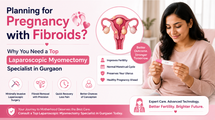
Planning for a baby is an exciting journey, but discovering you have uterine fibroids can introduce unexpected worries. For many women, non-cancerous muscular growths in the uterus can disrupt conception, complicate early pregnancy, or cause painful symptoms during gestation. When fertility preservation is your highest priority, consulting a Top Laparoscopic Myomectomy Specialist in Gurgaon is crucial. Advanced keyhole surgery offers a precise, uterus-sparing solution that removes problematic fibroids while optimizing your reproductive health.
How Uterine Fibroids Impact Fertility and Pregnancy
Uterine fibroids vary significantly in size, number, and location. Depending on where they grow, they can affect your reproductive system in several ways:
-
Submucosal Fibroids: These grow inside the uterine cavity, altering the shape of the uterus, blocking embryo implantation, or increasing early miscarriage risks.
-
Intramural Fibroids: Located within the muscular wall of the uterus, large intramural fibroids can block fallopian tubes or compress the uterine lining.
-
Subserosal Fibroids: Positioned on the outer wall, these may cause significant pelvic pressure, backache, or pain during intercourse.
Experiencing unexplained fertility delays or recurring pregnancy loss makes seeing a Top Laparoscopic Myomectomy Specialist in Gurgaon essential. A specialist evaluates whether fibroid removal is necessary to create a healthy environment for your future baby.
Why You Need a Top Laparoscopic Myomectomy Specialist in Gurgaon Before Conceiving
Navigating fibroids while planning a pregnancy requires expert surgical precision. You need a Top Laparoscopic Myomectomy Specialist in Gurgaon to preserve your uterine wall and optimize your fertility for several key reasons:
1. Delicate Uterine Wall Reconstruction
Removing a fibroid creates a defect in the uterine muscle that must be meticulously sutured. A skilled specialist ensures the uterine wall heals strongly enough to expand safely during future pregnancy and labor.
2. Minimally Invasive Keyhole Precision
Laparoscopic myomectomy uses tiny incisions, high-definition cameras, and specialized keyhole instruments. Partnering with a Top Laparoscopic Myomectomy Specialist in Gurgaon reduces internal scarring and pelvic adhesions that could otherwise cause fallopian tube blockages.
3. Faster Healing and Reduced Conception Delay
Compared to traditional open surgery, keyhole surgery reduces post-operative pain and infection risks. Healing quickly allows you to safely attempt pregnancy much sooner under your specialist’s guidance.
Key Signs to Consult a Top Laparoscopic Myomectomy Specialist in Gurgaon
Not all fibroids require surgical removal before conception. However, you should consult a Top Laparoscopic Myomectomy Specialist in Gurgaon if you notice any of the following indicators:
-
Infertility or Delayed Conception: Difficulty conceiving after months of trying, where fibroids distort the uterine cavity.
-
Recurrent Pregnancy Loss: A history of repeated miscarriages linked to submucosal or intramural growths.
-
Severe Heavy Period Bleeding: Heavy menstrual flow leading to iron-deficiency anemia and fatigue.
-
Persistent Pelvic Pain & Pressure: Continuous lower abdominal discomfort, frequent urination, or painful intercourse.
-
Rapidly Growing Fibroids: Scans showing accelerated growth that could interfere with fetal development.
Receiving a comprehensive evaluation from a Top Laparoscopic Myomectomy Specialist in Gurgaon gives you a clear, personalized timeline for surgery, recovery, and safely trying to conceive.
What to Expect During Your Surgical Consultation
When you consult a Top Laparoscopic Myomectomy Specialist in Gurgaon, your pre-conception care plan will follow a structured process:
-
Advanced Imaging Diagnostics: High-resolution 3D pelvic ultrasounds or pelvic MRIs to precisely map every fibroid’s location, size, and depth.
-
Customized Surgical Strategy: A step-by-step plan that removes fibroids while preserving healthy uterine tissues and fallopian tubes.
-
Clear Pre-Conception Guidance: Clear timelines on when it is safe to start trying for a pregnancy after complete uterine healing.
Working closely with a Top Laparoscopic Myomectomy Specialist in Gurgaon ensures that your path to motherhood is backed by expert clinical care, modern technology, and a dedicated uterine-preservation strategy.
Anatomical Classification and Biomechanical Impact of Uterine Fibroids
Uterine leiomyomas (fibroids) are benign monoclonal tumors arising from the smooth muscle cells of the myometrium. Their clinical manifestation, surgical difficulty, and effect on reproductive physiology depend heavily on their anatomical location relative to the uterine wall and endometrial cavity.
                  ┌────────────────────────────────────────┐
                  │   FIGO Subepithelial Classification    │
                  └───────────────────┬────────────────────┘
                                      │
         ┌────────────────────────────┼────────────────────────────┐
         ▼                            ▼                            ▼
 [Submucosal (Type 0-2)]      [Intramural (Type 3-5)]    [Subserosal (Type 6-7)]
 - Exophytic into cavity      - Within myometrium         - Projects outward
 - Causes heavy bleeding      - Expands uterine wall      - Causes pressure/bulk
 - Direct fertility barrier   - Impairs contractility     - Minimal cavity impact
The FIGO Classification System
The International Federation of Gynecology and Obstetrics (FIGO) categorizes fibroids into an 8-point system to guide surgical approach:
-
Type 0: Completely pedunculated submucosal fibroid within the endometrial cavity.
-
Type 1: Submucosal fibroid with $<50\%$ intramural extension.
-
Type 2: Submucosal fibroid with $\ge 50\%$ intramural extension.
-
Type 3: $100\%$ intramural fibroid abutting the endometrium without cavity distortion.
-
Type 4: Pure intramural fibroid residing entirely within the myometrium.
-
Type 5: Subserosal fibroid with $\ge 50\%$ intramural extension.
-
Type 6: Subserosal fibroid with $<50\%$ intramural extension.
-
Type 7: Pedunculated subserosal fibroid connected to the serosa by a vascular stalk.
-
Type 8: Transcervical, parasitic, or extrauterine fibroid location.
Impairment of Uterine Physiology
Fibroids alter pelvic biomechanics and reproductive capacity through distinct mechanisms:
-
Endometrial Receptivity Impairment: Submucosal (Types 0–2) and transmural fibroids cause localized endometrial ulceration, alter homeobox gene expression ($\text{HOXA10}$ and $\text{HOXA11}$), and induce local cytokine inflammation that disrupts embryo implantation.
-
Myometrial Hypercontractility: Intramural tumors disrupt orderly wave-like myometrial contractions during sperm transport and early blastocyst development.
-
Vascular Engorgement and Menorrhagia: Large intramural and subserosal fibroids compress pelvic venous plexuses, increasing overall uterine volume, dilating endometrial venules, and causing structural uterine bleeding.
Pre-Operative Mapping and Vascular Assessment Protocols
Precise anatomical localization prior to laparoscopic myomectomy prevents intraoperative conversion to open laparotomy and reduces blood loss.
| Diagnostic Tool | Clinical Parameters Evaluated | Surgical Planning Advantage |
| High-Definition 3D Pelvic Ultrasonography | Tumor number, precise diameter, location relative to main uterine vessels | Rapid screening and real-time mapping of localized anterior/posterior wall lesions |
| Saline Infusion Sonohysterography (SIS) | Endometrial cavity contour, sub-mucosal component distortion | Differentiates between hysteroscopic vs. laparoscopic myomectomy candidate lesions |
| Multi-Planar Pelvic MRI (T2-Weighted) | Exact pseudocapsule vascularity, distinction between leiomyoma and adenomyoma | Identifies deep intramural fibroids, vascular pedicles, and relation to ureteric pathways |
| Color Doppler Imaging | Peripheral vessel velocity, Resistance Index ($\text{RI}$), and Pulsatility Index ($\text{PI}$) | Predicts intraoperative vascular bleeding risks and suitability for temporary occlusion techniques |
Advanced Hemostatic Strategies in Laparoscopic Myomectomy
Controlling myometrial hemorrhage is critical during the excision of multiple or large intramural fibroids. Advanced minimally invasive techniques combine vascular occlusive maneuvers with targeted pharmacological agents.
                             [Intraoperative Hemostasis]
                                          │
                  ┌───────────────────────┴───────────────────────┐
                  ▼                                               ▼
     [Pharmacological Agents]                         [Mechanical/Vascular]
     - Intra-myometrial Vasopressin                   - Transient Uterine Artery Ligation
     - Tranexamic Acid Infusion                       - Temporary Pelvic Tourniquet
     - Topical Hemostatic Matrix                      - Targeted Bipolar Coagulation
Transient Uterine Artery Ligation (TUAL)
In cases involving large ($>7\text{ cm}$) or deep intramural lesions, temporary laparoscopic clipping or suture ligation of the uterine arteries at their origin from the internal iliac vascular trunk reduces arterial blood flow by up to $80\%$. This creates a bloodless surgical field during myomectomy while maintaining tissue viability through collateral ovarian vessels. Clips are removed following complete myometrial reconstruction.
Infiltration of Vasoconstrictive Agents
Intra-myometrial injection of diluted vasopressin ($0.05\text{–}0.1\text{ IU/mL}$) into the pseudocapsule creates localized blanched vasoconstriction. This hydrodissection opens the natural tissue cleavage plane between the fibroid capsule and healthy myometrium, allowing precise enucleation.
Topical Hemostatic Matrices and Sealants
Following tissue reconstruction, flowable gelatin-thrombin matrices or oxidized regenerated cellulose sheets are applied directly to the uterine suture line to establish localized hemostasis and prevent oozing from deep vascular channels.
Myometrial Reconstruction and Multilayered Suture Techniques
Restoring the structural integrity of the uterine wall is essential to prevent post-operative uterine rupture during subsequent pregnancies and labor.
                          [Deep Intramural Defect]
                                     │
      ┌──────────────────────────────┼──────────────────────────────┐
      ▼                              ▼                              ▼
[Layer 1: Endometrium/Deep]    [Layer 2: Myometrium]         [Layer 3: Serosal Layer]
- Continuous running suture    - Interrupted/Baseball        - Subcuticular Lembert suture
- Excludes mucosal lining      - Obliterates dead space      - Prevents adhesion formation
Tiered Layer Closure
-
Deep Myometrial Layer: Continuous running or interrupted $2\text{-}0$ barbed (V-Loc / Stratafix) or Vicryl sutures are placed to close the basal defect and eliminate internal hematoma formation.
-
Intermediate Myometrium: A second layer of continuous $1\text{-}0$ synthetic absorbable sutures re-approximates intermediate muscle fibers, distributing tensile strength evenly along the incision line.
-
Serosal Closure (Subcuticular Lembert): The outer serosa is inverted using an ultra-fine $3\text{-}0$ suture in a baseball or Lembert pattern. Inverting serosal edges minimizes raw tissue exposure, reducing pelvic adhesion formation.
Barbed Suture Innovations
Self-anchoring barbed sutures eliminate the need for manual laparoscopic knot tying under tension. This reduces operative time, maintains consistent tissue tension along the entire suture length, and minimizes warm ischemia duration.
In-Bag Contained Tissue Extraction (Morcellation Safety)
To prevent the intraperitoneal dissemination of undetected occult leiomyosarcomas or benign smooth muscle tissue (parasitic leiomyomas), laparoscopic myomectomy uses closed, in-bag containment systems.
┌─────────────────────────────────────────────────────────────────────────────┐
│                   In-Bag Power Morcellation Protocol                        │
├─────────────────────────────────────────────────────────────────────────────┤
│ 1. Fibroid Specimen Enucleated and Kept Intact Within Abdomen               │
│ 2. Heavy-Duty Endobag Introduced Through Expanded 10-12 mm Trocar           │
│ 3. Fibroid Placed Entirely Inside Sealable Endobag                          │
│ 4. Bag Aperture Exteriorized Through Abdominal/Umbilical Incision           │
│ 5. Pneumoperitoneum Established WITHIN the Sealed Containment Bag          │
│ 6. Morcellator Tip Inserted Under Direct Laparoscopic Visualization         │
│ 7. Tissue Fragments Retained Inside Bag; Interior Irrigation & Closure      │
└─────────────────────────────────────────────────────────────────────────────┘
By ensuring that electromechanical power morcellation occurs exclusively inside an impermeable containment bag, surgical teams eliminate cellular spillage into the peritoneal cavity while allowing large fibroids to be extracted safely through sub-centimeter abdominal incisions.
Adhesion Prevention Protocols Post-Myomectomy
Pelvic adhesions following uterine surgery can cause chronic pain, tubal factor infertility, and difficult re-operations. Preventing postoperative adhesions requires meticulous tissue handling and barrier agents.
-
Minimal Electrocautery Usage: Bipolar energy application is restricted to active bleeding vessels. Excessive thermal desiccation causes tissue necrosis, which triggers fibrin deposition and adhesion formation.
-
Intra-Peritoneal Barrier Gels: Hyaluronic acid-carboxymethylcellulose or cross-linked hyaluronan gels are applied over uterine suture lines to form a temporary physical barrier during the 5–7 day re-epithelialization window.
-
Peritoneal Washout: Thorough irrigation with warm heparinized ringer-lactate solution removes blood clots, cell debris, and inflammatory exudates before abdominal closure.
Post-Myomectomy Obstetric Management and Labor Pathways
Managing pregnancy following laparoscopic myomectomy requires balancing natural delivery options against the risk of uterine rupture.
                                  [Post-Myomectomy Conception]
                                               │
                      ┌────────────────────────┴────────────────────────┐
                      ▼                                                 ▼
          [Superficial/Subserosal Excision]             [Deep Intramural Cavity Penetration]
                      │                                                 │
                      ▼                                                 ▼
          [Trial of Labor After Myomectomy]                 [Elective Cesarean Section]
           (37-39 Weeks Full Term Delivery)                  (Planned at 37-38 Weeks Term)
Timing of Subsequent Conception
Patients are advised to wait 3 to 6 months following surgery before attempting pregnancy. This healing interval allows complete collagen deposition, myometrial remodeling, and re-vascularization of the uterine wall.
Mode of Delivery Determination
-
Elective Cesarean Delivery (37–38 Weeks): Mandatory if the myomectomy involved full-thickness myometrial incision entering the endometrial cavity, extensive reconstruction of deep intramural defects, or if post-operative wound healing was compromised by infection or hematoma.
-
Trial of Labor After Myomectomy (TOLAM): Considered safe for patients who underwent excision of superficial subserosal or small intramural fibroids where myometrial defect depth was minimal and suturing was completed without complication.
Comprehensive Reproductive and Surgical Care: Dr. Preeti Rastogi
When evaluating options for complex fibroid excision, minimal-access gynaecology, and fertility preservation, choosing an experienced surgical specialist is vital. Renowned as the best gynaecologist in gurugram, Dr. Preeti Rastogi offers world-class surgical expertise paired with compassionate, patient-centered care.
Qualifications and Clinical Leadership
-
Director & Head of Department: Leads Obstetrics and Gynaecology at Medanta – The Medicity, Gurugram, managing complex gynecological and high-risk surgical cases.
-
International Training & Credentials: Completed over a decade of advanced sub-specialty training in the United Kingdom, holding MRCOG (UK) and fellowships in Minimal Access Surgery, Urogynecology, and High-Risk Obstetrics from Singleton Hospital and the University Hospital of Wales, Cardiff.
-
Certified Da Vinci Xi Robotic Surgeon: Pioneer in applying robotic-assisted precision technology to myomectomy, total laparoscopic hysterectomy, and complex deep pelvic floor reconstruction.
-
Fertility-Focused Organ Preservation: Specializes in tissue-sparing, bloodless myomectomy protocols designed to preserve uterine integrity and optimize future pregnancy outcomes for women facing severe fibroids.
About Dr. Preeti Rastogi
-
Current Leadership Role: Director and Head of Department (HOD) in Obstetrics & Gynaecology at Medanta – The Medicity, Gurugram.
-
Extensive Clinical Experience: Over 25 to 30 years of total experience across top tertiary care hospitals in the UK (including Singleton Hospital, Swansea, and University Hospital of Wales, Cardiff) and India.
-
Educational Background: MBBS (G.R. Medical College, Gwalior), DNB in Obstetrics & Gynaecology (St. Stephen’s Hospital, Delhi), and MRCOG (Royal College of Obstetricians and Gynaecologists, UK).
-
Advanced Surgical Credentials: Certified Da Vinci Xi Robotic Surgeon specializing in minimally invasive laparoscopic myomectomy, hysteroscopy, and high-risk pregnancy care.
#laparoscopic myomectomy specialist #gurgaon #best fibroid surgeon #gurugram #minimally invasive fibroid removal #uterus sparing surgery #keyhole #myomectomy doctor #fibroids #pregnancy #fertility preserving #fibroid removal #submucosal fibroid surgery #intramural #myomectomy specialist #pre conception #fibroid treatment #robotic myomectomy doctor #advanced laparoscopic gynecologist #heavy menstrual bleeding #treatment #uterine fibroid specialist #gurgaon
FAQ
Q1: Do all uterine fibroids need to be removed before trying to get pregnant?
A: No, not all fibroids require surgical removal before pregnancy. Asymptomatic subserosal or small intramural fibroids that do not distort the uterine cavity often allow for normal conception and pregnancy. However, submucosal fibroids or large intramural fibroids that alter the uterine lining should be evaluated by a specialist, as they can hinder embryo implantation or increase miscarriage risks.
Q2: How does a laparoscopic myomectomy help preserve fertility in women with fibroids?
A: Laparoscopic myomectomy is a specialized, uterus-sparing procedure that precisely removes fibroids through keyhole incisions while keeping the rest of the uterus intact. By carefully suturing and reconstructing the uterine wall, the surgeon restores normal uterine shape, prevents pelvic adhesions, and preserves your natural ability to conceive and carry a child to full term.
Q3: Why consult Dr. Preeti Rastogi, best gynecologist in Gurgaon, when planning pregnancy with fibroids?
A: Dr. Preeti Rastogi, best gynecologist in Gurgaon, brings over 25 years of UK-trained clinical expertise as Director & HOD at Medanta – The Medicity. As a certified Da Vinci Xi robotic surgeon and expert in minimal access surgery, she specializes in complex, fertility-preserving myomectomies and high-risk obstetrics, ensuring maximum safety for both mother and future child.
Q4: How long should I wait to get pregnant after undergoing a laparoscopic myomectomy?
A: On average, gynecologists recommend waiting 3 to 6 months after a laparoscopic myomectomy before trying to conceive. This waiting period allows the reconstructed uterine muscle layer (myometrium) to heal fully, ensuring it can handle the physical stretch and strain of a growing pregnancy.
Q5: Will I need a C-section delivery if I get pregnant after a laparoscopic myomectomy?
A: The need for a C-section depends on the location and depth of the fibroids removed during surgery. If deep intramural fibroids required full-thickness suturing of the uterine wall, an elective Caesarean section is often recommended to prevent uterine rupture during labor. Your gynecologist will determine the safest delivery plan based on your surgical records.
Q6. How large can a fibroid be for safe laparoscopic removal?
A: With advanced laparoscopic techniques—such as transient uterine artery ligation, hydrodissection, and contained in-bag morcellation—fibroids exceeding $10\text{–}12\text{ cm}$ in diameter or multiple fibroids can be removed laparoscopically by an experienced minimal access surgeon. Pre-operative evaluation using MRI or 3D ultrasound determines whether laparoscopic or robotic-assisted surgery is most appropriate.
Q7. What is the difference between robotic-assisted myomectomy and traditional laparoscopic myomectomy?
A: Robotic myomectomy utilizes the Da Vinci Xi system, providing 3D high-definition visualization, 10x magnification, and wrist-articulated instruments that mimic human hand motions. This allows greater dexterity when suturing deep myometrial defects or operating in tight pelvic spaces. Standard laparoscopic surgery uses straight-line instruments and 2D/3D camera monitors. Both are minimally invasive options compared to open surgery.
Q8. How long is the recovery period following laparoscopic myomectomy?
A: Because laparoscopic myomectomy avoids large abdominal incisions, hospital stays are typically short ($24\text{–}48\text{ hours}$). Patients return to light daily activities within 7 to 10 days, with complete internal myometrial healing occurring within 4 to 6 weeks. This compares to a 6 to 8 week recovery timeframe for open abdominal surgery.
Q9. Will fibroids grow back after laparoscopic removal?
A: Laparoscopic myomectomy removes all visible and accessible fibroid nodules. However, because the underlying genetic or hormonal drivers remain, microscopic seedling cells can develop into new fibroids over time. Regular annual pelvic ultrasounds help monitor long-term myometrial health.
Q10. What makes Dr. Preeti Rastogi the best gynaecologist in gurugram for laparoscopic myomectomy?
A: Dr. Preeti Rastogi brings over 25 years of clinical experience, including 10+ years of UK tertiary hospital training. As Director and Head of Department at Medanta – The Medicity and a certified Da Vinci Xi Robotic Surgeon, her mastery of minimal access surgery, tissue-sparing reconstruction, and personalized fertility preservation establishes her as the best gynaecologist in gurugram.