Heavy Bleeding & Fibroids? When to Consult the Top Laparoscopic Myomectomy Doctor in Gurgaon.
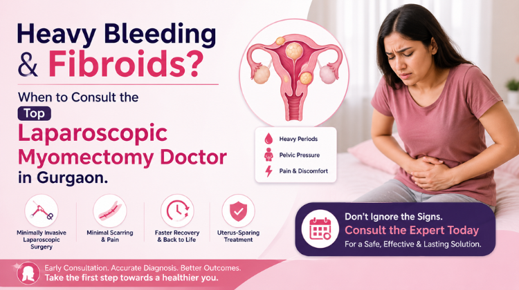
Dealing with unusually heavy periods, persistent pelvic pressure, and unexplainable fatigue can feel overwhelming. For many women, these disruptive symptoms stem from non-cancerous growths in the uterus known as uterine fibroids. When medications fail to provide relief, consulting a specialist becomes essential. Knowing when to reach out to the Top Laparoscopic Myomectomy Doctor in Gurgaon can mean the difference between prolonged suffering and a smooth, uterine-preserving recovery.
Understanding Uterine Fibroids and Heavy Bleeding
Uterine fibroids are benign muscular tumors that develop in or around the wall of the uterus. While some women experience no noticeable issues, others endure severe symptoms—most notably heavy menstrual bleeding (menorrhagia).
Excessive bleeding often results from fibroids that distort the uterine cavity or prevent the uterine muscles from contracting properly to stop blood flow. Over time, chronic blood loss leads to iron-deficiency anemia, causing constant exhaustion, dizziness, and shortness of breath. When conservative medical treatments fail to manage these symptoms, seeking guidance from the Top Laparoscopic Myomectomy Doctor in Gurgaon is the most effective next step toward long-term relief.
When Should You Consult the Top Laparoscopic Myomectomy Doctor in Gurgaon?
Knowing when to transition from home management or general gynecological visits to specialized surgical evaluation is crucial. You should book an appointment with the Top Laparoscopic Myomectomy Doctor in Gurgaon if you experience any of the following red flags:
-
Soaking Through Sanitary Products: Needing to change pads or tampons every hour for several consecutive hours.
-
Prolonged Periods: Menstrual bleeding that lasts longer than 7 to 8 days regularly.
-
Large Blood Clots: Consistently passing blood clots larger than a quarter during your period.
-
Symptoms of Anemia: Chronic fatigue, pale skin, weakness, or unexplained headaches due to low hemoglobin levels.
-
Pelvic Pain and Pressure: A constant sense of heaviness, fullness, or acute pain in your lower abdomen and back.
-
Fertility and Pregnancy Concerns: Difficulty conceiving or experiencing recurrent miscarriages linked to submucosal or intramural fibroids.
Why Choose Laparoscopic Myomectomy Over Traditional Surgery?
Laparoscopic myomectomy is a advanced, minimally invasive surgical procedure that allows surgeons to remove uterine fibroids while keeping the rest of the uterus intact. When performed by the Top Laparoscopic Myomectomy Doctor in Gurgaon, this procedure offers substantial benefits compared to open abdominal surgery:
-
Uterus Preservation: Unlike a hysterectomy, a laparoscopic myomectomy removes only the fibroids, keeping future pregnancy options open.
-
Minimal Inclusions and Scarring: Performed through tiny keyhole incisions, resulting in minimal cosmetic scarring.
-
Faster Recovery Time: Most patients return to normal daily activities within 2 to 3 weeks.
-
Reduced Post-Operative Pain: Significantly less pain and lower requirement for strong pain medications during recovery.
-
Lower Risk of Complications: Reduced risk of post-surgical infections and heavy blood loss.
Consulting the Top Laparoscopic Myomectomy Doctor in Gurgaon ensures that your surgery is planned using state-of-the-art imaging, precise keyhole instruments, and gentle tissue handling techniques.
What to Expect During Your Consultation with the Top Laparoscopic Myomectomy Doctor in Gurgaon
Your initial consultation with the Top Laparoscopic Myomectomy Doctor in Gurgaon is designed to map out a clear, individualized treatment path. During this visit, the specialist will:
-
Review your medical history, menstrual tracking logs, and prior treatments.
-
Perform a detailed physical and pelvic examination.
-
Order high-resolution diagnostic tests, such as pelvic ultrasounds or pelvic MRIs, to pinpoint the exact size, number, and location of your fibroids.
-
Evaluate your fertility goals to ensure the surgical plan prioritizes uterine wall integrity.
-
Explain the step-by-step surgical plan, expected recovery timelines, and post-operative care.
By partnering with the Top Laparoscopic Myomectomy Doctor in Gurgaon, you gain access to comprehensive care that prioritizes both your immediate symptom relief and long-term reproductive health.
Surgical Precision in Intramural, Cervical, and Broad Ligament Myomectomy
Laparoscopic myomectomy requires tailored anatomical strategies when dealing with atypical, deep, or giant uterine fibroids. While standard subserosal and superficial intramural lesions present straightforward operative planes, complex variants such as broad ligament, retroperitoneal, cervical, and deep intramural fibroids demand advanced surgical dexterity and deep knowledge of pelvic retroperitoneal anatomy.
┌─────────────────────────────────────────────────────────────────────────────┐
│                       Anatomical Challenges by Location                     │
├─────────────────────────────────────────────────────────────────────────────┤
│ 1. Broad Ligament Fibroids: Displace ureters and uterine artery trunks      │
│ 2. Cervical Fibroids: Distort pelvic floor anatomy and cervical canal      │
│ 3. Deep Intramural Fibroids: Border endometrial cavity; high vascularity    │
│ 4. Parasitic Fibroids: Receive blood supply from omentum/mesentery          │
└─────────────────────────────────────────────────────────────────────────────┘
Broad Ligament Fibroids
Fibroids originating between the leaves of the broad ligament alter normal pelvic geometry. As these tumors expand, they displace the uterine artery superiorly and push the ipsilateral ureter laterally or inferiorly. During laparoscopic excision:
-
Ureterolysis: The posterior leaf of the broad ligament is opened to identify and trace the path of the ureter from the pelvic brim down to the ureteric tunnel.
-
Controlled Dissection: The fibrous capsule of the broad ligament fibroid is incised on its anterior avascular surface. Traction and counter-traction are applied to enucleate the mass while preserving adjacent uterine vessels and the ureter.
Cervical Fibroids
Cervical leiomyomas create significant distortion of the lower uterine segment, displacing the bladder flap superiorly and compressing the rectum posteriorly.
-
Bladder Mobilization: The uterovesical peritoneum is sharply reflected downward to push the urinary bladder safely away from the anterior cervical wall.
-
Encapsular Enucleation: Incisions are strictly placed directly over the pseudocapsule to avoid injuring the ascending branches of the uterine artery running parallel to the lateral uterine border.
Parasitic and Extrauterine Leiomyomas
Parasitic fibroids detach from the main uterine corpus and establish secondary blood supplies from the omentum, mesentery, or retroperitoneal structures. Laparoscopic extraction requires systematic bipolar desiccation and division of feeding mesenteric vessels without compromising bowel perfusion.
Intraoperative Laparoscopic Ultrasound (LUS) for Deep-Seated Tumors
While preoperative high-resolution MRI provides a structural map of uterine fibroids, intraoperative shifts in tissue position during pneumoperitoneum can complicate the precise localization of small, deep intramural fibroids that do not distort the serosal contour.
          [Palpation Limitation in Laparoscopy]
                           │
                           ▼
          [Intraoperative Laparoscopic Ultrasound]
                           │
      ┌────────────────────┴────────────────────┐
      ▼                                         ▼
[Exact Depth Mapping]                [Cavity Intrusion Check]
- Identifies small intramural tumors  - Measures distance to endometrium
- Prevents unnecessary myometrial cuts - Prevents accidental mucosal breaches
High-Frequency Flexible Probes
A specialized 7.5–10 MHz flexible laparoscopic ultrasound probe is introduced through a 10 mm trocar port and placed directly against the uterine serosa. This enables real-time acoustic cross-sections of the myometrium.
Key Applications of Intraoperative LUS
-
Mapping Non-Palpable Fibroids: Laparoscopic instruments lack the direct tactile feedback of open laparotomy. Ultrasound imaging pinpoint small, deep-seated leiomyomas ($<2\text{ cm}$) embedded within the posterior or lateral uterine walls, preventing missed tumors.
-
Assessment of Endometrial Proximity: LUS accurately measures the thickness of the remaining myometrial bridge between the deep margin of the fibroid and the endometrial cavity, allowing the surgeon to plan incisions that minimize the risk of breaching the endometrial mucosa.
-
Evaluating Cleavage Planes: Acoustic shadow mapping helps identify dense calcifications or degenerative changes within the pseudocapsule, guiding the entry point for hydrodissection.
Uterine Fibroid Pseudocapsule Preservation and Neurovascular Integrity
The fibroid pseudocapsule is a thin, hypervascular, and neurogenic structure surrounding the leiomyoma. It is composed of compressed healthy myometrium, blood vessels, and a rich network of neuropeptides and nerve fibers (including substance P, vasoactive intestinal polypeptide, and neuropeptide Y).
  [Healthy Myometrium] ──> [Neurovascular Pseudocapsule] ──> [Fibroid Core]
                                   │
                    ┌──────────────┴──────────────┐
                    ▼                             ▼
         [Preserved Pseudocapsule]     [Aggressive Excision]
         - Promotes rapid healing       - Causes excess bleeding
         - Restores uterine nerves      - Increases scar tissue
         - Protects myometrial reserve  - Depletes healthy muscle
Physiological Advantages of Pseudocapsule Preservation
Traditional surgical approaches often excised the pseudocapsule along with the fibroid mass. Modern minimally invasive techniques prioritize preserving this surrounding capsule through intracapsular enucleation.
-
Neuro-Vascular Safeguarding: Preserving the pseudocapsule retains the underlying intrinsic nerve supply and micro-vasculature, accelerating post-operative tissue repair and muscular regeneration.
-
Enhanced Uterine Wound Healing: Intracapsular traction allows the surgeon to peel the fibroid directly out of its bed without tearing adjacent myometrial fibers. This reduces intraoperative tissue trauma, limits dead-space creation, and promotes uniform collagen remodeling.
-
Reduction of Post-Surgical Uterine Scarring: Maintaining functional myometrial thickness around the excision site preserves overall uterine compliance and contractility, which are critical for supporting full-term pregnancies.
Comprehensive Hemostatic Protocols: Pharmacological and Mechanical Interventions
Minimizing intraoperative blood loss during laparoscopic myomectomy is paramount for maintaining clear visual fields, preventing conversion to open surgery, and reducing the need for blood transfusions.
| Hemostatic Technique | Mechanism of Action | Operative Advantage | Clinical Considerations |
| Intramyometrial Vasopressin | Direct smooth muscle vascular constriction | Produces rapid, localized tissue blanching and hydrodissection | Must avoid direct intravascular injection; monitor blood pressure and heart rate |
| Transient Uterine Artery Ligation (TUAL) | Occludes main uterine arterial blood supply | Reduces total uterine blood inflow by up to 80% | Temporary procedure; ties or clips are removed post-reconstruction |
| Systemic Tranexamic Acid (TXA) | Antifibrinolytic; inhibits plasminogen activation | Decreases diffuse micro-vascular oozing from myometrial cuts | Administered intravenously prior to initial uterine incision |
| Pelvic Tourniquet Technique | Mechanical constriction of lower uterine segment | Temporarily cuts off blood flow during multi-fibroid extraction | Applied using a soft Penrose drain or suture loop around the cervix |
| Flowable Hemostatic Matrices | Thrombin-based local clotting cascade activation | Rapidly seals small non-suturable vascular beds | Applied directly into the deep surgical bed prior to closure |
                       [Comprehensive Hemostasis Pathway]
                                       │
            ┌──────────────────────────┴──────────────────────────┐
            ▼                                                     ▼
 [Systemic & Pre-Op Prep]                             [Intraoperative Control]
 - IV Tranexamic Acid (1g)                            - Intramyometrial Vasopressin
 - 3D Vascular Mapping                                - Transient Uterine Artery Clips
                                                      - Deep Barbed Layered Suturing
Hydrodissection and Vasoconstrictive Infiltration
A solution of vasopressin diluted in normal saline (0.05 to 0.1 units/mL) is injected directly into the subcapsular plane using a laparoscopic needle. This step achieves two clinical goals:
-
Physical Hydrodissection: The fluid volume creates fluid tension that separates the outer pseudocapsule from the inner tumor, clarifying the plane of dissection.
-
Potent Vasoconstriction: Vasopressin causes local constriction of small capillaries and arteriolar networks, turning an otherwise bloody dissection into a clean operative field.
Transient Uterine Artery Ligation (TUAL)
For solitary large intramural fibroids ($>8\text{ cm}$) or multiple deep intramural tumors ($>5\text{ nodules}$), temporary ligation of the uterine arteries offers excellent bleeding control.
-
The broad ligament’s posterior leaf is incised to expose the uterine artery as it branches from the internal iliac artery.
-
Vascular clips or temporary slip-knot sutures are applied to the bilateral uterine arteries.
-
Following complete fibroid enucleation and multi-layer suturing of the myometrium, the temporary clips or sutures are released, fully restoring blood flow to the uterus.
Biomechanical Principles of Myometrial Defect Closure and Scar Strength
Proper closure of the myometrial defect is the single most critical factor in preventing post-operative uterine rupture during subsequent pregnancies. Laparoscopic suturing must restore muscle alignment and eliminate internal voids where blood can collect.
                           [Myometrial Layer Closure]
                                       │
     ┌─────────────────────────────────┼─────────────────────────────────┐
     ▼                                 ▼                                 ▼
[Deep Base Layer]            [Intermediate Muscle Layer]        [Serosal Inversion Layer]
- 1-0 or 2-0 Barbed Suture   - Continuous 1-0 Suture            - 3-0 Subcuticular Lembert
- Closes deep dead space     - Realigns myometrial fibers       - Inverts tissue; stops adhesions
Layered Suturing Strategies
-
Deep Myometrial Reconstruction: Deep defects left by large intramural fibroids require multi-layered, continuous or interrupted suturing ($1\text{-}0$ or $2\text{-}0$ absorbable barbed suture). The first layer re-approximates the deepest muscle tissue adjacent to the endometrium, taking care not to pass through the endometrial mucosa itself.
-
Intermediate Muscle Re-alignment: Subsequent continuous or figure-of-eight layers close intermediate tissue spaces. Each pass of the needle should incorporate deep tissue bites to prevent purse-stringing or ischemic strangulation of healthy muscle.
-
Serosal Subcuticular Closure (Lembert Technique): The outer serosal edges are closed using an inverting Lembert or baseball suture technique with $3\text{-}0$ absorbable material. Inverting the raw serosal edges inward leaves a smooth uterine surface, significantly reducing the risk of bowel or omental adhesions.
Barbed vs. Standard Monofilament Sutures
The introduction of bidirectional and unidirectional barbed sutures (such as V-Loc or Stratafix) has transformed laparoscopic myomectomy:
┌─────────────────────────────────────────────────────────────────────────────┐
│                       Barbed vs. Standard Sutures                           │
├─────────────────────────────────────────────────────────────────────────────┤
│ Barbed Sutures:                                                             │
│  - Anchors along the entire length without knot tying                       │
│  - Maintains uniform, continuous tension along the surgical wound           │
│  - Reduces operative time and ischemic duration of tissues                  │
│                                                                             │
│ Standard Monofilament/Braided Sutures:                                      │
│  - Requires laparoscopic knot tying under variable tension                  │
│  - Tension concentrated solely at knot points, risking tissue tearing       │
└─────────────────────────────────────────────────────────────────────────────┘
Enclosed In-Bag Morcellation Protocols and Oncological Safety Standards
Power morcellation allows large fibroid specimens to be broken down into smaller tissue strips for removal through standard 10–12 mm trocar sites. However, uncontained morcellation carries a risk of spreading unrecognized leiomyosarcomas or benign tissue fragments (which can cause parasitic leiomyomas or endometriosis) throughout the peritoneal cavity.
┌─────────────────────────────────────────────────────────────────────────────┐
│                 Step-by-Step Enclosed In-Bag Morcellation                   │
├─────────────────────────────────────────────────────────────────────────────┤
│ Step 1: Complete Enucleation                                                │
│        The fibroid is fully detached and placed in the lower pelvis.        │
│                                                                             │
│ Step 2: Bag Deployment                                                      │
│        A heavy-duty specimen retrieval bag is introduced via a trocar.      │
│                                                                             │
│ Step 3: Specimen Capture                                                    │
│        The fibroid is placed inside the bag under direct camera view.       │
│                                                                             │
│ Step 4: Aperture Exteriorization                                            │
│        The mouth of the bag is pulled through the main umbilical port site. │
│                                                                             │
│ Step 5: Insufflation and Visualized Morcellation                            │
│        The bag interior is pneumatically insufflated; the morcellator       │
│        operates strictly within the sealed bag cavity.                      │
│                                                                             │
│ Step 6: Retrieval & Peritoneal Washout                                      │
│        The bag is sealed, removed, and the abdomen is irrigated.            │
└─────────────────────────────────────────────────────────────────────────────┘
Key Technical Principles for Contained Morcellation
-
Impermeable Multi-Layer Bags: Modern containment systems utilize tear-resistant, puncture-proof polyurethane bags designed to withstand electromechanical morcellator blade contact.
-
Dual-Port Visualization: A secondary laparoscope is introduced directly into the insufflated specimen bag alongside the morcellator blade. This setup maintains clear visualization of the blade’s position relative to the bag walls at all times.
-
Decontamination Protocol: Once tissue retrieval is complete, the bag is sealed and pulled through the umbilical incision. The pelvic cavity is then thoroughly irrigated with warm saline to ensure no residual cellular material remains.
Post-Operative Adhesion Mitigation and Pelvic Peritoneal Care
Pelvic adhesions following uterine surgery can bind the uterus to the bowel, bladder, or adnexa. This can cause chronic pelvic pain, painful intercourse, and mechanical tubal infertility. Minimizing scar tissue formation is a central priority during laparoscopic myomectomy.
                  [Adhesion Formation Prevention]
                                 │
     ┌───────────────────────────┼───────────────────────────┐
     ▼                           ▼                           ▼
[Surgical Precision]      [Physical Barriers]        [Peritoneal Hygiene]
- Minimal electrocautery  - Hyaluronic Acid Gels     - Warm saline lavages
- Inverting serosal cuts  - Oxidized Cellulose       - Thorough clot removal
Causes of Post-Myomectomy Adhesions
Adhesions develop when surgical trauma disrupts the peritoneal mesothelium, exposing raw subperitoneal connective tissue. Blood clots and inflammatory exudates collect on these un-epithelialized surfaces, forming fibrin bridges that mature into permanent fibrous scar bands within 5 to 7 days post-surgery.
Advanced Adhesion Reduction Strategies
-
Meticulous Hemostasis: Completely stopping blood oozing from the uterine suture lines eliminates the raw fibrin substrate needed for adhesion formation.
-
Thermal Energy Management: Limiting continuous monopolar cautery prevents widespread tissue drying and collateral thermal necrosis. Ultrasonic scalpels or precise bipolar forceps are used instead to keep tissue damage localized.
-
Resorbable Anti-Adhesion Barriers: Applying resorbable physical barriers—such as cross-linked hyaluronic acid gels or oxidized regenerated cellulose sheets—over uterine suture lines keeps surgical sites separated from adjacent peritoneal surfaces during the initial healing window.
-
Peritoneal Lavage: Rinsing the pelvis with warm, heparinized Ringer’s lactate solution removes microscopic blood clots and tissue debris before closing the abdomen.
Obstetric Care Pathways: Fertility, Pregnancy, and Mode of Delivery
Preserving future reproductive potential is a primary goal when performing laparoscopic myomectomy for women of childbearing age. Managing post-operative healing and planning subsequent pregnancies requires a structured clinical strategy.
                                [Conception Pathway Post-Myomectomy]
                                                 │
                        ┌────────────────────────┴────────────────────────┐
                        ▼                                                 ▼
            [Superficial/Pedunculated Removal]                [Deep Intramural Cavity Ingress]
                        │                                                 │
                        ▼                                                 ▼
            [Wait 3 Months Before Trying]                     [Wait 6 Months Before Trying]
                        │                                                 │
                        ▼                                                 ▼
            [Trial of Labor After Myomectomy]                 [Elective Cesarean Delivery]
             (Full term 38-40 Weeks Delivery)                  (Planned Delivery at 37-38 Weeks)
Timing of Subsequent Conception
-
Superficial Intramural or Subserosal Myomectomy: Patients can typically begin attempting pregnancy 3 months post-surgery, provided routine ultrasound evaluation shows complete healing of the myometrium.
-
Deep Intramural or Extensive Multi-Fibroid Reconstruction: A healing phase of 6 months is recommended to allow for full collagen maturation and restoration of tensile strength in the uterine wall.
Scar Evaluation and Uterine Monitoring
Pre-conception 3D pelvic ultrasonography or contrast sonohysterography is used to confirm that the uterine wall has healed properly and that the endometrial contour remains uniform. During pregnancy, high-resolution ultrasounds monitor myometrial thickness at the previous operative sites.
Mode of Delivery Determination
The decision between a trial of labor after myomectomy (TOLAM) and an elective cesarean section depends on several intraoperative factors:
-
Indications for Elective Cesarean Section (37–38 Weeks):
-
Full-thickness incision that entered the endometrial cavity during fibroid removal.
-
Extensive reconstruction required for large or multiple deep intramural fibroids.
-
Complications during healing, such as post-operative hematoma formation within the uterine wall.
-
History of electrosurgical tissue desiccation near the uterine defect.
-
-
Candidates for Trial of Labor (TOLAM):
-
Excision limited to pedunculated subserosal or shallow intramural fibroids.
-
Intact, un-breached deep myometrium with a smooth, well-healed outer serosa.
-
Unremarkable post-operative recovery with no signs of uterine scar defect on follow-up imaging.
-
World-Class Surgical Care in Gurugram: Dr. Preeti Rastogi
Choosing the right minimal-access surgeon is essential when seeking treatment for complex uterine fibroids, pelvic organ preservation, and fertility protection. Known as the best gynaecologist in gurugram, Dr. Preeti Rastogi brings over 25 years of specialized international clinical experience to complex women’s healthcare.
┌─────────────────────────────────────────────────────────────────────────────┐
│                 Dr. Preeti Rastogi: Key Clinical Profile                    │
├─────────────────────────────────────────────────────────────────────────────┤
│ • Leadership: Director & Head of Department, Obstetrics & Gynaecology      │
│   at Medanta – The Medicity, Gurugram                                  │
│ • UK Training: Over 10 years of senior experience in UK NHS hospitals,       │
│   including Singleton Hospital (Swansea) & University Hospital of Wales     │
│   (Cardiff)                                                           │
│ • Qualifications: MBBS, DNB (Obs & Gynae), MRCOG (London, UK)                │
│ • Fellowships: Minimal Access Surgery, Urogynecology, and High-Risk         │
│   Obstetrics (UK)                                                            │
│ • Robotic Expertise: Certified Da Vinci Xi Robotic Surgeon specializing     │
│   in myomectomy, hysterectomy, and pelvic floor reconstruction             │
└─────────────────────────────────────────────────────────────────────────────┘
Expert Surgical Approach
Dr. Preeti Rastogi focuses on organ-preserving, bloodless myomectomy techniques designed to protect uterine integrity and support future pregnancy goals. Her mastery of advanced laparoscopic and Da Vinci Xi robotic surgery ensures precise tumor removal, minimal blood loss, rapid recovery, and excellent cosmetic outcomes.
Comprehensive Gynecological Services
Beyond laparoscopic myomectomy, Dr. Rastogi offers specialized treatment across a wide spectrum of complex conditions:
-
Minimal Access & Robotic Surgery: Laparoscopic hysterectomy, deep excision of severe stage 4 endometriosis, and ovarian cystectomy.
-
High-Risk Obstetrics: Comprehensive management of complex pregnancies, including severe pre-eclampsia, gestational diabetes, twin gestations, and previous uterine scars, with a strong commitment to supporting safe vaginal deliveries.
-
Urogynecology & Pelvic Floor Disorders: Advanced treatment for pelvic organ prolapse, stress urinary incontinence (TVT/TOT slings), and custom pelvic reconstructive procedures.
-
Reproductive Medicine & Menstrual Disorders: Comprehensive diagnostic evaluations for fibroid-induced heavy bleeding, adenomyosis, dysmenorrhea, and reproductive failure.
Her patient-centered approach, dedication to evidence-based clinical care, and high surgical success rates solidify her reputation as the best gynaecologist in gurugram.
Taking the First Step Toward Recovery
Heavy bleeding and fibroid pain do not have to dictate your daily life. Modern minimally invasive surgical techniques make fibroid removal safer, faster, and more effective than ever. If heavy periods are affecting your health or fertility, scheduling an evaluation with the Top Laparoscopic Myomectomy Doctor in Gurgaon will help you regain control over your well-being.
Facts About Dr. Preeti Rastogi
-
Current Position: Senior Director & Head of Department (HOD) in Obstetrics & Gynaecology at Medanta – The Medicity, Gurugram.
-
Experience: Over 25 to 30 years of extensive clinical experience across top tertiary care hospitals in the United Kingdom (including Singleton Hospital, Swansea, and University Hospital of Wales, Cardiff) and India.
-
Education & Credentials: MBBS (G.R. Medical College, Gwalior), DNB in Obstetrics & Gynaecology (St. Stephen’s Hospital, Delhi), and MRCOG (Royal College of Obstetricians and Gynaecologists, UK).
-
Specializations: Advanced Laparoscopic Myomectomy, Certified Da Vinci Xi Robotic Surgery, High-Risk Obstetrics, and Urogynaecology.
FAQ
Q1: What causes heavy bleeding in women with uterine fibroids?
A: Heavy menstrual bleeding (menorrhagia) caused by uterine fibroids occurs when these non-cancerous muscular growths increase the inner surface area of the uterine lining (endometrium). Submucosal and intramural fibroids also disrupt normal uterine muscle contractions, preventing blood vessels from clamping shut during menstruation, which leads to heavy flow and large blood clots. Heavy Bleeding & Fibroids? When to Consult the Top Laparoscopic Myomectomy Doctor in Gurgaon.
Q2: What is the difference between a hysterectomy and a laparoscopic myomectomy for fibroids?
A: A hysterectomy is the complete surgical removal of the entire uterus, which permanently eliminates periods and the ability to bear children. In contrast, a laparoscopic myomectomy is a minimally invasive procedure that surgically removes only the fibroids while reconstructing and preserving the uterus, making it the preferred option for women who wish to preserve their fertility. Heavy Bleeding & Fibroids? When to Consult the Top Laparoscopic Myomectomy Doctor in Gurgaon.
Q3: How do I know if I need to see Dr. Preeti Rastogi, best gynecologist in Gurgaon, for my fibroids?
A: You should consult Dr. Preeti Rastogi, best gynecologist in Gurgaon, if you experience severe symptoms such as periods lasting longer than a week, passing large blood clots, soaking through pads every hour, severe pelvic pressure, or difficulty getting pregnant due to fibroids. Her UK-trained expertise in advanced laparoscopic and robotic myomectomy ensures precision-driven, uterus-preserving care. Heavy Bleeding & Fibroids? When to Consult the Top Laparoscopic Myomectomy Doctor in Gurgaon.
Q4: What is the average recovery time after a laparoscopic myomectomy?
A: Most patients undergoing a laparoscopic myomectomy spend 1 to 2 days in the hospital for observation. Initial recovery allows for light walking within 24 to 48 hours, and most women fully return to work and daily light routine activities within 2 to 3 weeks, which is significantly faster than the 6-to-8-week recovery required for traditional open abdominal surgery. Heavy Bleeding & Fibroids? When to Consult the Top Laparoscopic Myomectomy Doctor in Gurgaon.
Q5: Can fibroids return after being removed by laparoscopic myomectomy?
A: While a laparoscopic myomectomy permanently removes existing fibroids, new fibroids can potentially develop over time from microscopic muscle cells. The risk depends on age, genetics, hormonal factors, and the total number of fibroids removed. Regular follow-up appointments with your gynecologist help monitor and manage any future risk. Heavy Bleeding & Fibroids? When to Consult the Top Laparoscopic Myomectomy Doctor in Gurgaon.
Q6. How does a surgeon decide between laparoscopic myomectomy and open myomectomy?
A: The choice between laparoscopic and open surgery depends on fibroid size, number, depth within the uterine wall, and location relative to major pelvic blood vessels. Advances in laparoscopic suturing, hydrodissection, and contained morcellation allow experienced minimally invasive surgeons to safely remove large ($>10\text{ cm}$) or multiple fibroids laparoscopically. However, if an abdominal scan shows severe pelvic adhesions or a very high number of intramural fibroids ($>10\text{–}12\text{ nodules}$) distributed throughout the uterus, open surgery may be recommended to ensure complete reconstruction.
Q7. Will removing my fibroids laparoscopically improve my chances of getting pregnant?
A: Yes, particularly if the fibroids distort the endometrial cavity (FIGO Types 0, 1, 2) or block the tubal ostia. Removing intramural and submucosal fibroids restores normal uterine shape, improves blood flow to the endometrium, reduces chronic inflammation, and eliminates physical barriers to implantation. Many patients conceive naturally or achieve successful outcomes through assisted reproductive techniques (such as IVF) within 6 to 12 months following surgery.
Q8. What is the typical hospital stay and recovery timeline for laparoscopic myomectomy?
A: Because laparoscopic myomectomy relies on small sub-centimeter abdominal incisions rather than a large open incision, post-operative pain is minimal and recovery is fast. Most patients are discharged within 24 to 48 hours of surgery. Light daily activities can usually be resumed within 7 to 10 days, and full physical activity—including exercise and routine work—typically resumes within 3 to 4 weeks.
Q9. Are there risks associated with power morcellation during laparoscopic fibroid removal?
A: Uncontained power morcellation carries a risk of spreading undetected uterine sarcomas or benign tissue fragments throughout the abdominal cavity. Modern surgical practice mitigates this risk by performing all morcellation inside a sealed, impermeable containment bag (enclosed in-bag morcellation). This approach contains all tissue fragments during breakdown, ensuring safe specimen extraction through small incision sites without compromising oncological safety.
Q10. Why is Dr. Preeti Rastogi considered the best gynaecologist in gurugram for complex fibroid surgery?
A: Dr. Preeti Rastogi brings over 25 years of international clinical experience, including more than a decade of senior practice in top UK NHS hospitals. As Director and Head of Department at Medanta – The Medicity and a certified Da Vinci Xi Robotic Surgeon, her specialized expertise in tissue-sparing myomectomy, advanced layered suturing, and fertility preservation makes her the best gynaecologist in gurugram for managing complex uterine fibroids.